
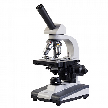
Микромед 1 вар. 1−20 обеспечивает возможность вывода изображения в режиме реального времени на экран ПК с помощью видеоокуляра (в комплект не входит). Видеоокуляр устанавливается в тубус микроскопа вместо окуляра.
Микроскоп рассчитан на длину тубуса 160 мм, объективы стандарта DIN, парфокальная высота объективов 45 мм.
Комплектность
Составные части:
Штатив (со встроенным в основание осветителем с галогенной лампой 20Вт) — 1Револьвер на 4 позиции объективов — 1 — Установлен на штативеНасадка монокулярная поворотная на 360º с наклоном на 45º - 1Столик прямоугольный механический (125×130 мм) двухкоординатный (70×30 мм) — 1 — Установлен на штативеСменные части:
Центрируемый Конденсор Аббе светлого поля А1,25 регулируемый по высоте с держателем светофильтров — 1 — Установлен на штативеКонденсор темного поля иммерсионный, А 1,36−1,25 — 1 — Поставляется по доп. заказуКонденсор темного поля сухой, А 0,9 -1 — Поставляется по доп. заказуОбъектив-ахромат 4х/0,1 160/0,17 — 1Объектив-ахромат 10х/0,25 160/0,17 — 1Объектив-ахромат 20х/0,4 160/0,17 — 1 — Поставляется по доп. заказуОбъектив-ахромат 40х/0,65 160/0,17 (подпружиненный) — 1Объектив-ахромат 60х/0,85 160/0,17 (подпружиненный) — 1 — Поставляется по доп. заказуОбъектив-ахромат 100х/1,25 ми 160/0,17 (подпружиненный) — 1Окуляр 10х/18 с центроуказателем — 1 — Установлен в тубус насадкиОкуляр 10х/18 с сеткой — 1 — Поставляется по доп. заказуОкуляр 10х/18 со шкалой — 1 — Поставляется по доп. заказуОкуляр 10х/18 с перекрестием — 1 — Поставляется по доп. заказуОкуляр 5х/18 — 1 — Поставляется по доп. заказуОкуляр 12,5х/15 — 1 — Поставляется по доп. заказуОкуляр 16х/15 - 1 — Поставляется по доп. заказуОкуляр 20х/11 - 1 — Поставляется по доп. заказуВидеоокуляр — 1 — Поставляется по доп. заказуСветофильтр голубой — 1Светофильтр зеленый — 1Светофильтр желтый — 1Светофильтр матовый — 1Принадлежности и запасные частиШнур сетевой — 1 — Установлен в основаниеЧехол — 1Флакон с иммерсионным маслом — 1Лампа галогенная 20Вт цоколь G4 — 2 — Одна установлена в штативе микроскопаВставка плавкая — 2 — Одна установлена в штативе микроскопаОтвертка (или ключ-шестигранник для крепления визуальной насадки) — 1Руководство по эксплуатации — 1Отличительные особенностиПо сравнению с простейшими учебными моделями Микромед 1 вар. 1−20 имеет:
- Двукоординатный предметный столик с коаксиальными рукоятками.
- Коаксиальный механизм грубой и точной фокусировки.
- Механизм фокусировки с регулировкой жесткости хода грубой фокусировки.
- Широкопольные окуляры с удаленным зрачком.
- Источник света — галогенная лампа комфортной для глаз цветовой температурой, с регулировкой яркости, что позволяет комфортно работать с объективами всех увеличений.
- Микроскоп Микромед 1 выпускается в трех вариантах — монокулярный, бинокулярный и тринокулярный. Данная монокулярная модель — идеальный инструмент для быстрых лабораторных работ в ВУЗах, где за одним микроскопом работают по очереди несколько человек (не требуется перенастройка визуальной насадки), визуальная насадка поворотная на 360º для более удобной совместной работы.
- Широкий ассортимент дополнительного оборудования увеличивает возможности микроскопа.


